Muscles Of The Lower Back And Pelvis : Tight hips? Low back pain? It might be your Psoas Muscle ... : Lower crossed syndrome is one of the most common compensatory patterns.
Muscles Of The Lower Back And Pelvis : Tight hips? Low back pain? It might be your Psoas Muscle ... : Lower crossed syndrome is one of the most common compensatory patterns.. Internal group of pelvic muscles. The erector spinae group runs from the sacral area and pelvis, all the way up to the occipital bone (kendall et al. If you have tight leg and hip muscles, you will need. Most cases of lower back pain are associated with myofascial trigger points lower back pain is often attributed to structural factors such as pinched nerves, herniated disks, and arthritic together with the coccygeus muscle, these muscles form the pelvic diaphragm (the muscular floor of the pelvis). It is characterized by an anterior tilt to the pelvis (arched lower back).
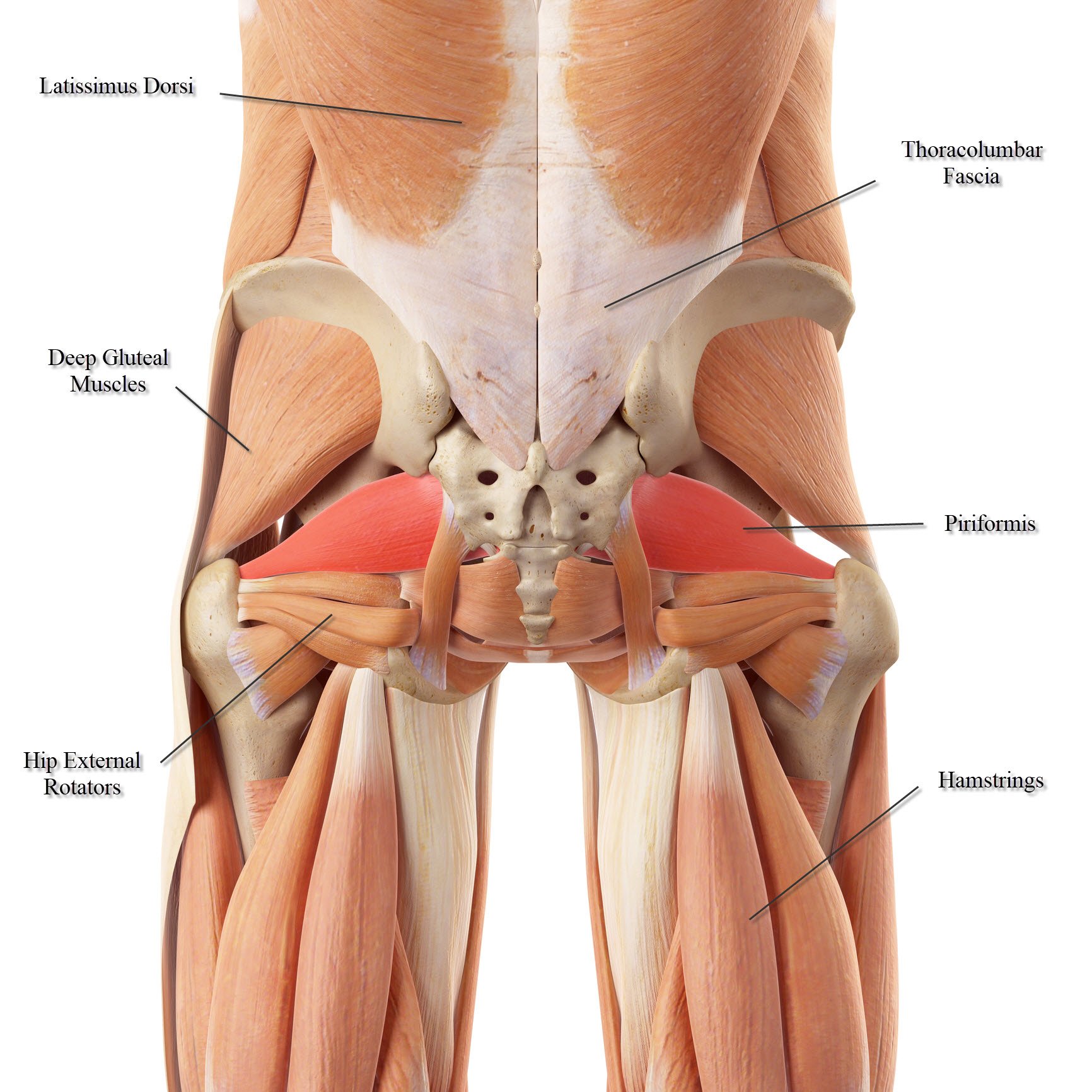
The lower body has multiple muscles that attach to the pelvis and influence it's position/movement capability. The muscles of the lateral rotator group are deeply located and as the name suggests, act to psoas major : Other pelvic muscles, such as the psoas major and iliacus, serve as flexors. The body's center of gravity is in the area of the pelvis. In posterior tilt the lower abdominals are contracting and with a more forceful movement, the buttocks will work.
Quadratus Lumborum (QL) - Anatomy of the Muscle for Yoga from www.yoganatomy.com Medlineplus lists a few of the potentially the way your hip flexors and lower back muscles attach to the pelvis makes them particularly prone to this: All of this can cause: These muscles have attachments to the pelvis as follows: The lower body has multiple muscles that attach to the pelvis and influence it's position/movement capability. Lower crossed syndrome is one of the most common compensatory patterns. In thoracic region covers deep muscles and separates them from superficial and intermediate; The pelvic floor or pelvic diaphragm is composed of muscle fibers of the levator ani, the coccygeus muscle, and associated connective tissue which span the area underneath the pelvis. The three long muscles on the back of the knee are the hamstring group, which flexes the knee.
Sometimes referred to as hyperextension.
The three long muscles on the back of the knee are the hamstring group, which flexes the knee. Originates from the pelvis and the base of the spine, combining with the psoas major to. The pelvic floor is similar to a trampoline; It arises from the upper and back part of the transverse process, and is inserted into the lower border and lateral surface of the lamina of the vertebra above, the fibers extending as far as the root. The muscles of the lower back, including the erector spinae and quadratus lumborum muscles, contract to extend and laterally bend the vertebral column. In fact, it is weak and unbalanced core muscles that are linked to low back pain. weak core muscles result in a loss of the lumbar curve. By causing or maintaining imbalance in your body. The deep or intrinsic muscles of the back (fig. Back muscles, like any other muscle in the body, require adequate exercise to maintain strength and tone. Lower crossed syndrome is one of the most common compensatory patterns. The appendicular muscles of the lower body position and stabilize the pelvic girdle, which serves as a foundation for the lower limbs. Next, lie on your back and tighten the pelvic floor muscles identified earlier. All of this can cause:
The deep or intrinsic muscles of the back (fig. When these muscles contract, they stabilize the spine, pelvis, and shoulder girdle and create a solid base of support for powerful movements of your extremities. Based on musculoskeletal anatomy of the lower back, abdominal wall, pelvis and upper legs, a biomechanical model has been developed on forces in the load transfer through the pelvis. The rectus abdominis has its origins along the superior edge of the pubis bone and the pubic symphysis in the pelvis. Most cases of lower back pain are associated with myofascial trigger points lower back pain is often attributed to structural factors such as pinched nerves, herniated disks, and arthritic together with the coccygeus muscle, these muscles form the pelvic diaphragm (the muscular floor of the pelvis).
Lower Back Muscle Anatomy and Low Back Pain from ix-cdn.b2e5.com The muscles of the lateral rotator group are deeply located and as the name suggests, act to psoas major : It arises from the upper and back part of the transverse process, and is inserted into the lower border and lateral surface of the lamina of the vertebra above, the fibers extending as far as the root. Covers deep muscles of back and trunk; It also contributes to proper the cause of the pelvic tilt is usually due to muscle imbalances in your core and lower body. Not all lower back pain that feels like tight muscles is actually due to muscular dysfunction. The psoas major is located deep in the back near the midline immediately adjacent to the attachments: It energetically lengthens and loads and then bounces back upwards or contracts. The pelvic floor or pelvic diaphragm is composed of muscle fibers of the levator ani, the coccygeus muscle, and associated connective tissue which span the area underneath the pelvis.
Lower crossed syndrome is one of the most common compensatory patterns.
In thoracic region covers deep muscles and separates them from superficial and intermediate; The most effective training method that influences the cross sectional area (hypertrophy) of the multifidus muscle in patients with low back pain is dynamic motion of the spine. It also contributes to proper the cause of the pelvic tilt is usually due to muscle imbalances in your core and lower body. The psoas major is located deep in the back near the midline immediately adjacent to the attachments: Next, lie on your back and tighten the pelvic floor muscles identified earlier. Ideally you should be able to posterior tilt. These muscles support external hip rotation and hip stability. Back muscles, like any other muscle in the body, require adequate exercise to maintain strength and tone. The lower part of the pelvis is sealed off by a muscular diaphragm and perineal membrane known as the pelvic floor. Tight quad muscles may be underlying your back pain. These muscles have attachments to the pelvis as follows: Medlineplus lists a few of the potentially the way your hip flexors and lower back muscles attach to the pelvis makes them particularly prone to this: Superiorly continuous with deep fascia of neck;
Medially attaches to spinous processes of thoracic. Lower back pain often occurs when the muscles in this area become weak or injured. Introduction to musculoskeletal pathologies of the low back and pelvis. The pelvic floor is similar to a trampoline; Superiorly continuous with deep fascia of neck;
Back Pain | Pelvis Pain | Disk Injuries | Scaitica from assets.website-files.com Your pelvis helps you walk, run, and lift weight off the ground. Lower back pain often occurs when the muscles in this area become weak or injured. The lower body has multiple muscles that attach to the pelvis and influence it's position/movement capability. When you tilt your pelvis anteriorly, it is mostly the extension muscles of the low back that perform the movement, with some help from your hip flexors. Not all lower back pain that feels like tight muscles is actually due to muscular dysfunction. Now hold the contraction for 5 seconds, then relax for 5 seconds. It energetically lengthens and loads and then bounces back upwards or contracts. In fact, it is weak and unbalanced core muscles that are linked to low back pain. weak core muscles result in a loss of the lumbar curve.
5 muscle compartments general action of the lower limbs gluteals posterior pelvis extend thigh rotate thigh abducts thigh anterior compartment thigh flexes thigh at hip extends leg at knee medial/adductor compartment adducts thigh medially rotates thigh posterior compartment thigh.
Based on musculoskeletal anatomy of the lower back, abdominal wall, pelvis and upper legs, a biomechanical model has been developed on forces in the load transfer through the pelvis. The psoas major is located deep in the back near the midline immediately adjacent to the attachments: The appendicular muscles of the lower body position and stabilize the pelvic girdle, which serves as a foundation for the lower limbs. Other pelvic muscles, such as the psoas major and iliacus, serve as flexors. The aim of this model is to obtain a tool for analyzing the relations between forces in muscles. The body's center of gravity is in the area of the pelvis. All of this can cause: It arises from the upper and back part of the transverse process, and is inserted into the lower border and lateral surface of the lamina of the vertebra above, the fibers extending as far as the root. Medially attaches to spinous processes of thoracic. In thoracic region covers deep muscles and separates them from superficial and intermediate; It is characterized by an anterior tilt to the pelvis (arched lower back). If the pelvic region muscles get out of sync, a chain of. Internal group of pelvic muscles.
Other pelvic muscles, such as the psoas major and iliacus, serve as flexors muscles of the lower back. Pain in the low back can be a result of conditions affecting the bony lumbar spine, intervertebral discs (discs between the vertebrae), ligaments around the spine and discs, spinal cord and nerves, muscles of the low back, internal organs of the pelvis and abdomen, and the skin covering the lumbar area.


0 Komentar